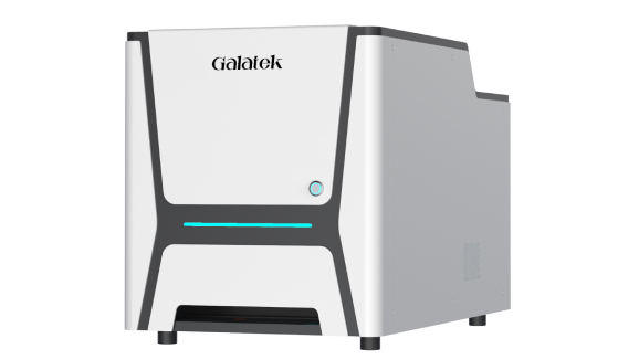
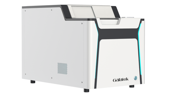
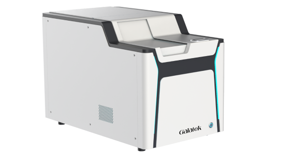
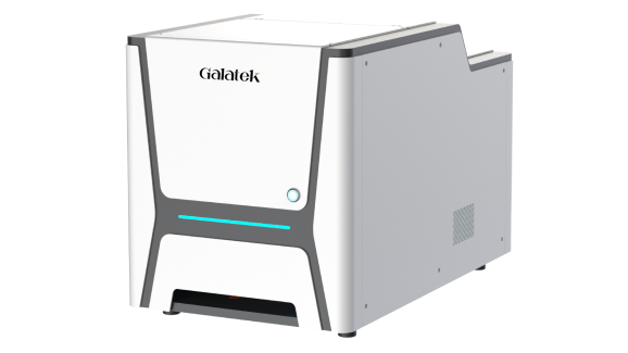
High Content Cell Imaging System_Galatek

Perspecta High Content Cell Imaging
Widefield and confocal imagers for 2D & 3D biology research
·

Solutions

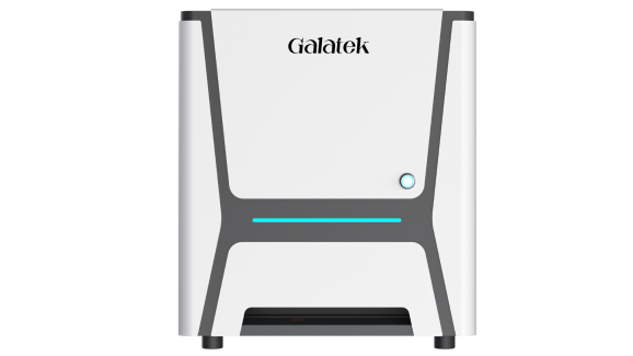
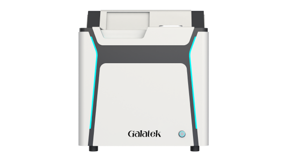
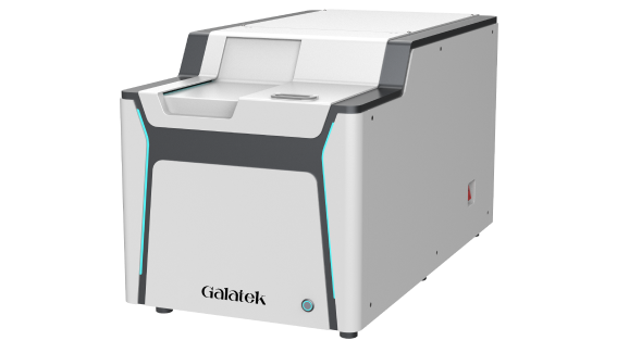
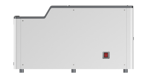
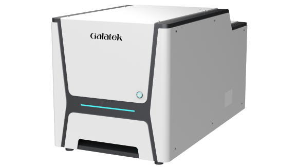
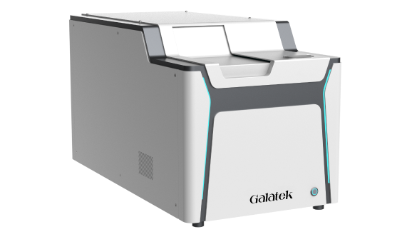
Perspecta High Content Cell Imager

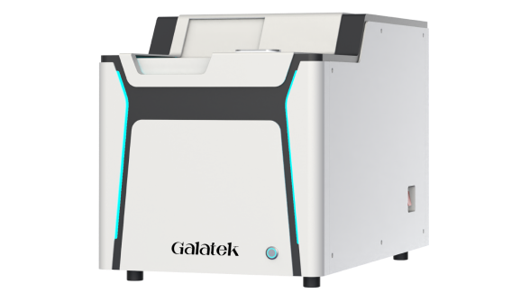
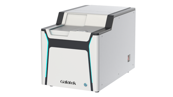
Perspecta Cx High Content Cell Imager

Perspecta High Content Cell Imager

The Perspecta High Content Cell Imager delivers high-speed, automated imaging across bright-field, fluorescence, and 3D samples.

Key capabilities:
• Supports diverse imaging modalities, including 3D, bright-field, and fluorescence
• Maintains stable environmental conditions for long-term live-cell observation
• Enables high-throughput imaging to accelerate screening and discovery workflows
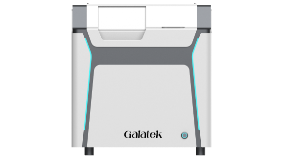
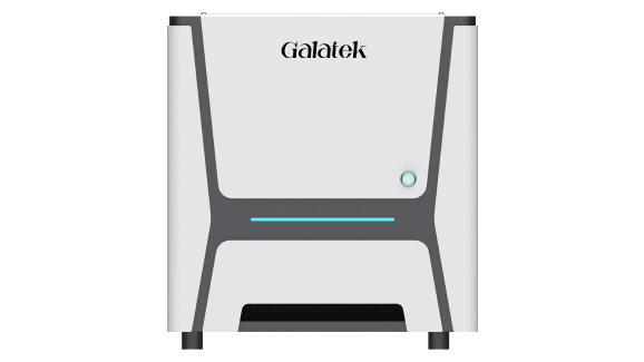
Perspecta Cx High Content Cell Imager

The PerspectaCx High Content Imager delivers high-speed, automated confocal imaging with optical sectioning for sharp, high-resolution 3D structural detail.

Key Capabilities:  
• Combines confocal and widefield imaging in one switchable system
• Reduces out of focus background for sharper, high-resolution 3D structural detail
• Integrates with automation and data management platforms for high-throughput 3D screening

Features

Flexible Configuration on Demand
Provides personalized configurations across brightfield, fluorescence, and 3D imaging to match experimental needs
Fast Imaging and Processing
A high-resolution CMOS camera (up to 31 fps) enables scanning of 384-well plates in under 30 minutes
High Imaging Quality
A high-energy LED light source paired with a 12 MP CMOS camera enhances signal strength for sensitive, high-quality imaging
Precise Environmental Control
Accurate temperature, humidity, and CO₂ control creates an ideal environment for long-term live-cell imaging
Seamless Upgrade to High-throughput Solutions
Integrates with Galatek's Galatek automation platform for unattended, high-throughput screening workflows

Product Parameter


Product Name

Perspecta High Content Cell Imager

Imaging Modes

Brightfield, color brightfield, fluorescence

Camera

CMOS camera

Pixel Size

1.85 μm

Camera Resolution

1200 MP

Camera Frame Rate

31 fps

Illumination

Brightfield (1 LED), fluorescence (4 LED +1 laser)

Fluorescence Channels (nm)

405, 488, 561, 638, 730 (Customized)

Objective Magnification

20x NA0.8, options for 4x /10x /40x 

Stage Precision

0.4 μm

Imaging Methods

Single-color, multicolor, time-lapse, Z-stacking, 2D deconvolution

Autofocus Methods

Image-based / 850 nm laser autofocus

Sample Containers

6-1,536 well-plates, slides, culture dishes

Control Rod

Focus knob, XY joystick, XYZ sensitivity adjustment buttons (6 buttons)

Software

Image capture and analysis

Application Scenarios

Brightfield Imaging
Brightfield Imaging
Cellular Fluorescence
Tissue Sections
Tissue Sections
Bioluminescence
Bioluminescence
Cell Painting
Cell Painting
Spatial Omics
Spatial Omics
Cell Tracking
Cell Tracking
High Content Imaging
Live Cell Imaging
Live Cell Imaging
Drug Screening
Drug Screening

Features

Switchable Optical System
Shift between confocal and widefield acquisition to match sample complexity – from routine 2D to structurally complex 3D and organoid samples
Fast Imaging and Processing
A high-speed sCMOS camera (up to 110 fps) enables simultaneous acquisition and analysis of full-plate confocal z-stack scans in under 2.5 minutes
Precise Environmental Control
Accurate temperature, humidity, and CO2 control creates an ideal environment for long-term live cell and organoid observation and imaging
High Imaging Quality
A 50 μm pinhole spinning-disk confocal paired with a 26 MP sCMOS camera reduces out-of-focus signal for sharper structural detail and clearer 3D reconstructions
Seamless Upgrade to High-throughput Solutions
Integrates with lab automation platforms for unattended, high-throughput 3D screening workflows

Product Parameter


Basic Parameters

Perspecta Cx High Content Imaging System

Optical system

Confocal imaging, widefield imaging (switchable)

Imaging modes

Brightfield, color brightfield, fluorescence, digital phase contrast, live cell dynamic imaging, Z-stack scanning

Imaging methods

50μm pinhole spinning disk confocal

Fluorescent channels

5 channels (405 nm, 488 nm, 561 nm, 638 nm, 730 nm)

Light source

Brightfield LED, fluorescence laser

Camera

sCMOS

Camera resolution

6224 × 4168 (26 MP)

Pixel size

3.76 μm

Camera frame rate

Up to 110 fps

Objective magnification

4-position motorized turret, 2x – 60x

(apochromatic or long working distance objectives are optional)

Stage travel

80 mm × 120 mm × 6 mm (X/Y/Z)

Stage repeatability

X/Y 0.4 μm; Z 0.1 μm

Focusing modes

Infrared laser focusing, image focusing

Consumable types

6 – 1,536 well plates, culture dishes, slides

Dimensions

820 mm x 445 mm x 500 mm

Operating environment

Temperature 5°C – 40°C; humidity ≤ 80%

Live cell culture module

Supports temperature, humidity, CO₂ concentration (optional)

Integration with laboratory data management systems

API available

Application Scenarios

Brightfield Imaging
Brightfield Imaging
Cellular Fluorescence
Tissue Sections
Tissue Sections
Bioluminescence
Bioluminescence
Cell Painting
Cell Painting
Spatial Omics
Spatial Omics
Cell Tracking
Cell Tracking
High Content Imaging
Live Cell Imaging
Live Cell Imaging
Drug Screening
Drug Screening

Contact Us

Have read and accepted the Privacy Policy and Terms of Service
Download document
Have read and accepted the Privacy Policy and Terms of Service